CN
  • Home
  • News
    • 2024
    • 2023
    • 2022
    • 2021
    • 2020
    • before
  • Members
    • Faculties
    • Postdocs
    • Ph.D Students
    • Master Students
    • undergraduate studen...
    • Alumni
  • Research
    • Biomedical Polymers ...
    • Gradient Biomaterial...
    • Interaction between ...
    • Self-Adaptive Biomat...
  • Publications
    • Publications
    • Books
    • Patents
    • Awards
  • Instruments
    • Instruments
    • Mini-Library
  • Recruitments

News

  • 2024

  • 2023

  • 2022

  • 2021

  • 2020

  • before

homepage  News  before
  • Nanotubes Protruding from Microcapsules: Phenomenon, mechanism and its applications

    2012-02-13

    Smart capsule systems are of high attraction due to their ability to respond to the alteration of environment conditions. But the morphology evolution of the microcapsules with the changes of the environment is very uncommon. Recently smart capsules which could reversibly respond to redox stimuli were fabricated in our group. In this process an interesting phenomenon of one-dimension nanostructures protruding from microcapsules were discovered and comprehensive studies have been performed to explore the mechanism and its applications. In preliminary studies, a strategy was applied to introduce ferrocene to the CaCO3 microparticles doped with poly(allylamine hydrochloride) (PAH) to fabricate microcapsules, which exhibit reversible responsibility to redox stimuli(Langmuir, 2011, 27(4): 1286-1291.). Through a similar strategy, pyrenecarboxaldehyde(Py-CHO) was reacted with the doped PAH to fabricate the PAH-Py microcapsules. Protrusion of one-dimensional nanotubes (1D-NT) or nanorods (1D-NR) from the PAH-Py microcapsules was discovered when the microcapsules were incubated in acidic solutions. And the protrusion process of the nanostructures was triggered by the environment conditions(ACS Nano, 2011, 5(5): 3930-3936.). Moreover, Attempt was then paid to control the 1D-NR growth state from the PAH-Py microcapsules by chemical cross-linking and surface modification(Journal of Materials Chemistry, 2012, 22: 2855-2858.), and the Py-CHO NR with multiple functions could be fabricated to obtain physical and chemical nanostructures(Chemistry of Materials, 2011, 23 (21): 4741-4747.). The above serious results are helpful for the development of a new class of methods to prepare multiple micro-nano structure capsules, which promotes the applications of capsules in the fields such as cell bionic technology, sensors, dynamic constitutional chemistry and so on. TEM images showing the process of nanotube protruding fromthe PAH-Pymicrocapsules incubated in pH 0 HCl for 0, 24, 48, 96, and 144 h, respectively. (f) Optical images (inset, a higher magnification) showing the protruded nanotubes fromthe PAH-Pymicrocapsules incubated in pH 0 HCl for 30 h. SEM images of amicrocapsulewith nanotubes after treatment in pH 0 HCl for (g) 30 h and (h) 72 h, respectively. (i) SEMimage of amicrocapsule with nanorods after treatment in pH 2 HCl for 1 h.

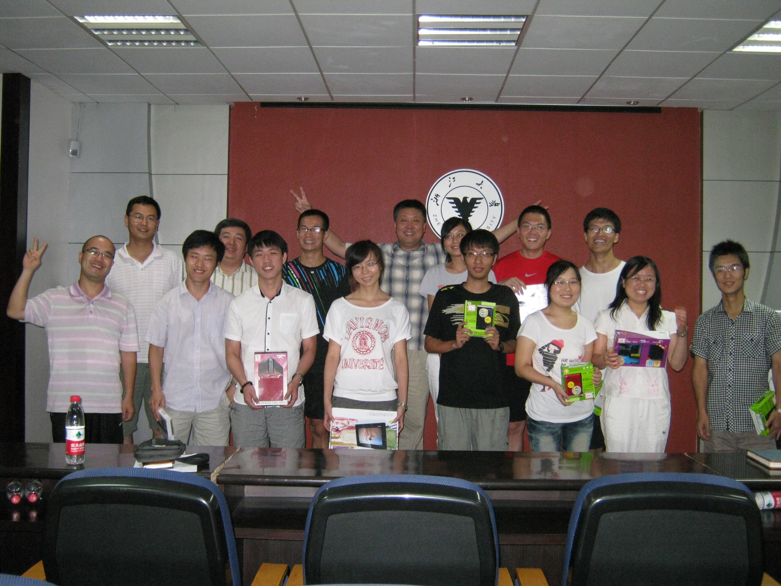
  • The 2011-2012 fall and winter semester summary in the group

    2012-02-12

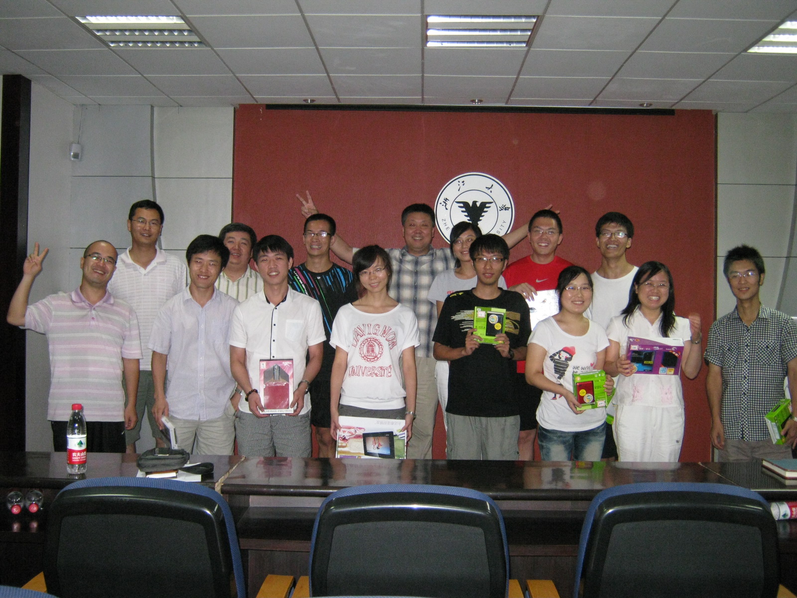
  • The 2011-2012 fall and winter semester summary in the group

    2012-02-12

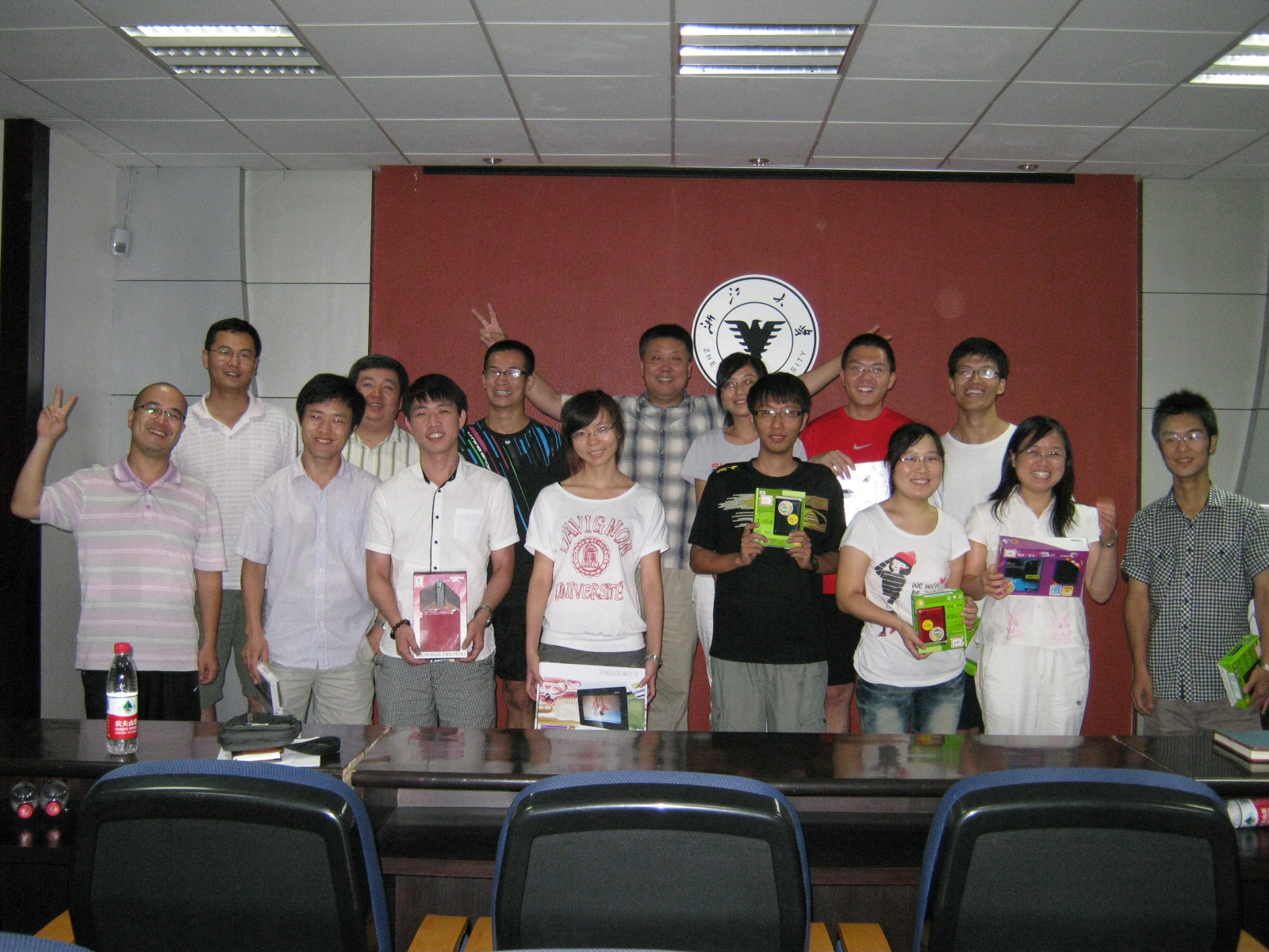
  • The 2011-2012 fall and winter semester summary in the group

    2012-02-12

  • The 2011-2012 fall and winter semester summary in the group

    2012-02-12

  • Chitosan Microcapsules With Micrometer-Thick and Layered Wall Structure by Stepwise Core-Mediated Precipitation

    2012-01-06

    Incubation of CaCO3 microparticles in chitosan (CS) solution at pH 5.2 and following with ethylenediaminetetraacetic acid disodium salt (EDTA) treatment resulted inCS single component microcapsules with an ultra-thick wall structure. Repeating the incubation causedstepwise increase of wall thickness and finally resulted in CS microcapsules with a layered structure. This unique method is mediated by precipitation of CS on the CaCO3 particles as a result of pH increase caused by the partial dissolution of CaCO3. The obtained CS capsules arestable at neutral pH.   Reference: Fabrication of Chitosan Single-Component Microcapsules With a Micrometer-Thick and Layered Wall Structure by Stepwise Core-Mediated Precipitation. Yuanyuan Han,Weijun Tong,* Yuying Zhang, Changyou Gao*.Macromolecular Rapid Communication, DOI =10.1002/marc.201100685.

  • IFBM2012-1st Circular

    2012-01-06

  • Controlling the migration behaviors of VSMCs by mPEG brushes of different molecular weight and density

    2012-01-06

    Cell migration is an important biological activity. Regulating the migration of vascular smooth muscle cells (VSMCs) is critical in tissue engineering and therapy of cardiovascular disease. In this work, methoxy poly(ethylene glycol)(mPEG) brushes of different molecular weight (Mw 2 kDa, 5 kDa and 10 kDa) and grafting mass (0-859 ng/cm(2)) were prepared on aldehyde-activated glass slides, and were characterized by X-ray photoelectron spectrometer (XPS) and quartz crystal microbalance with dissipation (QCM-d). Adhesion and migration processes of VSMCs were studied as a function of different mPEG Mw and grafting density. We found that these events were mainly regulated by the grafting mass of mPEG regardless of mPEG Mw and grafting density. The VSMCs migrated on the surfaces randomly without a preferential direction. Their migration rates increased initially and then decreased along with the increase of mPEG grafting mass. The fastest rates (∼24 μm/h) appeared on the mPEG brushes with grafting mass of 300-500 ng/cm(2) depending on the Mw. Cell adhesion strength, arrangement of cytoskeleton, and gene and protein expression levels of adhesion related proteins were studied to unveil the intrinsic mechanism. It was found that the cell-substrate interaction controlled the cell mobility, and the highest migration rate was achieved on the surfaces with appropriate adhesion force.    Jindan Wu, Zhengwei Mao*, Changyou Gao*, Controlling the migration behaviors of vascular smooth muscle cells by methoxy poly(ethylene glycol) brushes of different molecular weight and density. Biomaterials 2012; 33(3):810-20. Download PDF

  • The 2010-2011 spring and summer semester summary in the group

    2011-08-01

  • The 2010-2011 spring and summer semester summary in the group

    2011-08-01

  • The 2010-2011 spring and summer semester summary in the group

    2011-08-01

  • The 2010-2011 spring and summer semester summary in the group

    2011-08-01

  • Tug of war in the department

    2011-04-08


  • Tug of war in the department

    2011-04-08


  • per page 14 records  total 82 records 
  • firstpage <<previouspage nextpage>> endpage
  • PageNumber 4/6 jumpto 

Contact US

Phone:+86-0571-87951108
Email:cygao@zju.edu.cn
Address:Zhejiang University (Zijingang Campus) Hetong 6 Building 338, Hangzhou 310027, China 

Quick Links

浙江大学高分子科学与工程学系 博慧生物 滨江研究院 绍兴研究院 浙江大学
Copyright ©2024 Zhejiang University Biomedical Polymers for Tissue Repair and Regeneration Technical Support : Hzccx   Admin Login You are the 1000th visitor of this site